


Treating a Herniated Disc with Neural Therapy, Manual Medicine, and Magnetic Field Therapy Is…

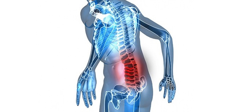
In whom is a herniated disc more commonly seen? A herniated disc — known in medical terminology as disc herniation — occurs in the population at a fairly high rate, roughly one in ten. It is most commonly seen between the ages of 35 and 50, occurring equally in both sexes. Due to spinal structure, taller individuals are at greater risk of developing a herniated disc. Those who perform heavy labor, homemakers, desk workers who must sit for long periods, drivers, professionals who stand constantly such as teachers, pharmacists, and waiters, those who work outdoors, and managers under intense stress are among the groups most likely to develop a herniated disc.
How does it occur?
A herniated disc occurs when the elastic cartilage tissue called the disc, located between the vertebrae, is pushed forward by pressure from the vertebrae, protrudes from the spinal cord sheath, and compresses the nerves that travel to various parts of the leg. Sometimes even a sudden strain, lifting something heavy, an awkward movement, or exposing the lower back to cold can cause a herniated disc. But the most important cause of a herniated disc is excessive tension in the muscles following prolonged stress and tightness.
What are the symptoms?
Pain radiating into one or both legs, numbness in the feet, restricted movement, and difficulty walking or sitting are symptoms of a herniated disc. If the condition progresses, symptoms such as impotence, quick fatigue, urinary incontinence, imbalance, and inability to walk may also occur.
How is a herniated disc diagnosed?
With today’s modern diagnostic methods, computed tomography and magnetic resonance imaging make it easy to diagnose a herniated disc and determine its severity.
How is herniated disc surgery performed?
With the microsurgical method used for herniated discs, patients can stand up and walk six or seven hours after surgery, and can go home after one night in the hospital. Thanks to this method, an incision of only 1.5–2 centimeters is made, and no stitches are needed on the skin surface after surgery.
Performing the surgery under advanced microscopes allows the nerves in the surgical area to be seen at 25 or 40 times magnification, thereby reducing the risk of nerve damage to zero.
 If the herniated disc has just begun
Treatment of a herniated disc depends on the degree to which the herniation — that is, the pressure exerted by the elastic disc material on the nerves running to the leg — has progressed. If there is only lower back and leg pain, with no numbness, loss of strength, or restricted movement, the herniated disc is in its early stage. In this case, the patient is advised to take muscle relaxants, rest in bed, and avoid movements that strain the lower back.
Recommendations for the patient
1) The patient must absolutely not lift weights exceeding one or two kilograms.
2) Bending forward or sideways, and bending the lower back, are prohibited. If something needs to be picked up from the floor, the patient should be told to squat down to pick it up.
3) Patients are advised to place a cushion behind the lower back while sitting, in a way that fills the lumbar curve, and not to sit for more than twenty minutes. If the patient’s occupation requires sitting for long periods, they are advised to walk every twenty minutes. Those who drive for long periods are advised to park their car every twenty minutes and walk around it a few times.
4) The patient is prohibited from reaching upward. If something needs to be taken from above, they should be told to use a stool or step up onto a stair.
5) The patient is reminded to always keep the lower back warm and to avoid drafts.
6) Patients with lower back and leg pain must absolutely avoid stress. Besides increasing pain, stress can also cause the herniated disc to progress.
7) The patient is advised to rest in bed during the time spent at home. Contrary to popular belief, extremely hard surfaces are more harmful. It is more appropriate to sleep on a good-quality spring mattress, in whatever position the patient finds most comfortable.
If the condition is at an advanced stage
If the patient’s complaints persist despite the above recommendations, rest, and muscle relaxants, physical therapy should be applied.
Classification of lower back pain
In the past, LBP (Low Back Pain) lasting longer than 6 months was considered chronic. Now, however;
• LBP lasting 0-4 weeks is considered acute
• LBP lasting 4-12 weeks is considered subacute
• LBP lasting longer than 12 weeks is considered chronic
• Manipulative therapy and massage are ineffective in unqualified hands
• Traction therapy is no longer recommended
• Evidence regarding acupuncture is insufficient (it is described as relieving pain but not curing it!)
• Biofeedback and TENS are ineffective on their own. They may be used in combined treatments!
• TP injections, prolotherapy, and Botox injections: conflicting results
• Epidural steroid injection is insufficient for radicular pain. Controlled studies are insufficient
• Cognitive therapy
Whereas Neural Therapy & Manual Medicine & Magnetic Field Therapy, combined with Acupuncture, is highly effective.
NEURAL THERAPY:
Treatment begins with an examination of the Adler-Langer points. Segments found positive by the Kibler skin rolling test are identified. Local treatment: local application of 1% lidocaine and procaine is performed to the locally affected areas.
Segmental treatment: application is made to irritation points within the segment, particularly to the trigger points of the gastrocnemius and soleus muscles. A quaddle is administered between L1-S5. L2 block and/or sacral canal (canalis sacralis) application.
If the patient has constipation or bowel problems, these should be treated.
Candida overgrowth in the intestines causes toxins to accumulate in connective tissue.
This should be investigated and identified using Reviqunat-Proqunat or Vega testing to find the source of the problem.
Any disturbance field (interference field) must absolutely be regulated. Unilateral tendinitis and arthritis are usually caused by a disturbance field.
The lower extremity circulation protocol is the treatment to be performed first. Disturbance fields within the same segment should be tested kinesiologically, and treatment should be planned according to the response obtained.
The patient should be reassessed at every visit, and necessary changes should be made to the treatment plan. In chronic cases, care must be taken not to fall into a classical viewpoint or routine.
Situations requiring surgery for a herniated disc
If the patient’s pain continues despite physical therapy and complementary medicine methods, or if there is an irreversible loss of strength, thinning of the leg, unbearable pain, and the CT or MRI scans show that a fragment has broken off from the disc, the solution is surgical intervention. This rate is below 5% of all herniated discs worldwide. In other words, surgery is the last resort and should not be used widely.
Sources Consulted:
• Nazlikul, H: Neural Therapy Textbook
• Nazlikul, H: Neural Therapy – Another Treatment Is Possible
• H. Barop’s Neural Therapy Atlas (Translator: H. Nazlikul)
• L. Fischer’s Neural Therapy Book (Translators: H. Nazlikul and Y. Tamam)
• James W. McNabb, Joint and Soft Tissue Injections (Translators: H. Nazlikul and Y. Tamam)
• Weinschenk, S: Neuraltherapie
• Fischer, L et al.: Lehrbuch Integrative Schmerztherapie