


What Should Be Done for Shoulder Pain? Neural Therapy for Shoulder-Related Movement Restriction,…

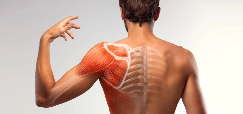
The shoulder joint is the most complex joint complex in the body. It consists of the glenohumeral, acromioclavicular, sternoclavicular, and scapulothoracic joints. For this reason, it would be more accurate to use the term shoulder complex rather than shoulder joint. The glenohumeral joint is a synovial, ball-and-socket type joint. Because the humeral head is large and the glenoid fossa is shallow, it has very little bony support. For this reason, joint stability is provided by soft structures such as the capsule, ligaments, and muscles. This also leads to a greater range of motion.
The static factors that limit the movement of the glenohumeral joint and provide stability are the joint surfaces, ligaments, joint capsule, intra-articular pressure, and labrum; the dynamic factors are the muscles crossing the joint.
The joint capsule, glenohumeral ligaments, and labrum are located around the glenoid fossa. The upper portion of the labrum merges with the tendon of the long head of the biceps. The labrum increases the depth of the glenoid by 50%.
The capsule is one of the important static stabilizers of the glenohumeral joint.
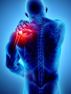 The glenohumeral ligaments (superior, middle, and inferior) are closely related to the capsule and reinforce it anteriorly. The inferior portion is the thickest and most important static stabilizer among all the glenohumeral ligaments. The coracohumeral ligament reinforces the anterosuperior surface of the capsule.
The subacromial (subdeltoid) bursa is located between the coracoacromial ligament, the acromion, and the supraspinatus tendon, and extends toward the deltoid muscle and beneath the coracoid process. The main function of this bursa is to reduce friction between the structures located in this region during movement, particularly between the humerus and the supraspinatus tendon.
Clinically, the most important structure in this region is the coracoacromial arch. This arch consists of the coracoid process of the scapula, the acromion, and the coracoacromial ligament connecting them. In the space beneath the arch lie the tendons of the rotator cuff muscles and the subacromial (subdeltoid) bursa.
The most important superficial muscles of the shoulder are the pectoralis major, deltoid, and trapezius muscles.
The deep muscle layer, on the other hand, is formed by the rotator cuff muscle group and the biceps brachii. The subscapularis muscle attaches to the lesser tubercle. The supraspinatus, infraspinatus, and teres minor muscles attach to the greater tubercle.
The serratus anterior, rhomboids, levator scapulae, and latissimus dorsi are trunk muscles that assist in stabilizing and moving the shoulder.
The superficial and deep structures of the shoulder joint are quite rich in nerve innervation. The nerve fibers originate from C4, C5, C6, and C7. Innervation of the ligaments, capsule, and synovial membrane is provided by the axillary, suprascapular, subscapular, and musculocutaneous nerves. These structures are also among the sites with the densest vegetative nervous system innervation.
The shoulder joint is also quite rich in arterial and venous circulation. However, the so-called "critical zone" near the attachment point of the supraspinatus tendon is an area with reduced vascularity. The main factor triggering changes related to the supraspinatus tendon is hypoxia in this region.
Lymphatic drainage of the upper extremity begins from the lymphatic vessels in the fingers and the lymphatic plexus in the skin of the hand. The flow is directed toward the forearm and arm. Part of it passes through the cubital nodes at the elbow and the deltopectoral nodes at the shoulder, draining into the axillary lymph nodes. These follow the neurovascular bundles and receive lymph from the joint capsule, periosteum, tendons, nerves, and muscles.
 When evaluating a patient presenting with shoulder complaints, we must first determine whether the problem is a local shoulder pathology. We know that, thanks to the communication between the structures within a segment via the spinal cord, no problem can remain purely local.
10% of people experience one or more episodes of shoulder pain and/or stiffness during their lifetime. These problems may stem from pathologies of the shoulder region itself, from other systemic diseases, or from distant factors.
When taking the patient's history, we should try to establish the temporal connection. A careful physical examination of the shoulder joint should then be performed, and any scar tissue, vaccination scars, or discoloration within the shoulder segment should be investigated. Positive findings obtained during the examination suggest that the problem originates from the shoulder and its segment. If there are none, the issue does not originate from the shoulder. The problem may be an interference field (IF); from this perspective, the Adler points should be examined. The intestines should also not be forgotten as a potential interference field. Trigger points that could cause shoulder pain should be examined. Neighboring joints should also not be overlooked, because we know that no joint becomes diseased on its own. The patient is reassessed for these possible pathologies. On the other hand, the patient is also evaluated locally. The problem may originate from the joint itself, or it may be a consequence of thoracic blockage. In this respect, complaints that worsen particularly at night can be considered a gold standard indicator.
The shoulder joint is also a region with many reflex zones. For this reason, previous tonsil infections or tonsillectomy, gallbladder-related complaints and operations, dental treatments the patient has undergone, and various cardiac problems present in the patient should be questioned and evaluated for temporal association. As is known, the shoulder region is a reflex zone for these organs.
Special tests performed during the physical examination play an important role in the differential diagnosis of shoulder pain. These tests mainly include:
- Painful arc test
- Supraspinatus test
- Neer test
- Hawkins test
- Drop arm test
- Infraspinatus test
- Subscapularis test
- Yergason test
- Speed test
 Treatment:
Our approach to treating shoulder disorders should be multidisciplinary. It is very difficult to achieve success in shoulder disorders when the neural therapy component is missing. Sometimes, in chronic cases, psychological support may be needed because the patient's shoulder use is restricted. Neural therapy has as important a place in treatment as these other branches—if not more so. Neural therapy is used successfully in acute shoulder injuries that do not require surgery.
Shoulder problems usually arise as a result of overload and frequently repeated strain. In the acute period, rest, analgesics and NSAIDs, and cold application are preferred; in the subacute and chronic periods, physical therapy agents and exercise are preferred.
Acute pathologies respond very well to neural therapy. The number of patients who go on to surgery is quite low. In chronic cases resistant to treatment, the usual causes are interference fields, thoracic blockage, trigger points, pathologies of neighboring joints, and, rarely, psychological overload. In chronic cases, it is possible to achieve success with repeated injections.
The initial approach in neural therapy should be to perform quaddles at painful and sensitive points and in the shoulder segment. The sites where the capsule and tendons—which are rich in VNS innervation—attach to bone should be treated in particular. Quaddles are performed on the C4-C7 segment. If no response is obtained, the problem lies higher up.
For this reason, it is necessary to make use of the effect of neural therapy in order to regulate the shoulder region.
Sources I Have Drawn On:
• Nazlikul, H: Neural Therapy Textbook
• Nazlikul, H: Neural Therapy, Another Treatment Is Possible
• H. Barop's Atlas of Neural Therapy (Translator: H. Nazlikul)
• L. Fischer's Neural Therapy Book (Translators: H. Nazlikul and Y. Tamam)
• James W. McNabb, Joint and Soft Tissue Injections (Translators: H. Nazlikul and Y. Tamam)
• Weinschenk, S: Neuraltherapie
• Fischer, L et al.: Lehrbuch Integrative Schmerztherapie