


Neural Therapy in Soft Tissue Trauma of the Foot and Ankle Definition: A sprain generally seen in…

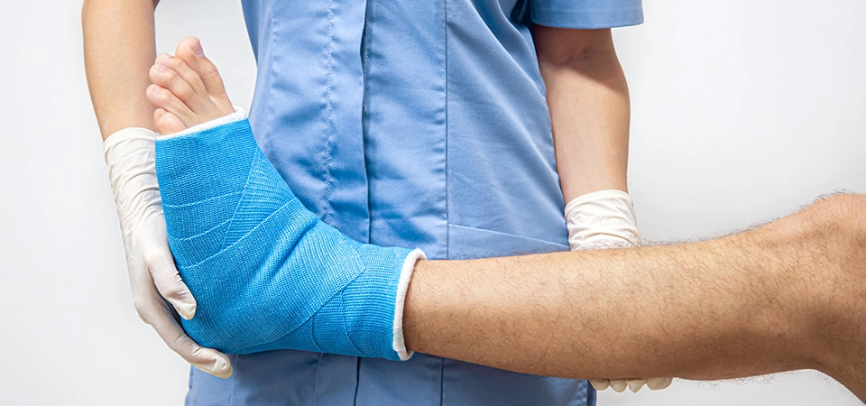
Definition: This is a sprain generally seen in the young, physically active population, resulting from mechanical loading of the soft tissues of the foot and ankle beyond what they can tolerate. The most commonly sprained ligaments are those on the lateral side of the ankle. The anterior talofibular ligament is most frequently involved. It is a serious public health issue, occurring at a rate of 1/10,000 per day. In approximately 90% of cases, a sprain occurs when the ankle is subjected to an inversion strain while in plantar flexion or supination. This is a situation frequently encountered in volleyball, basketball and football.
Findings Detected in the Patient:
Anterior Drawer Test: Shows laxity of the anterior talofibular ligament. Anterior displacement of the talus indicates a positive test.
Talar Tilt Test: A test that shows injuries to the anterior talofibular and calcaneofibular ligaments.
Syndesmotic rupture: A tear or injury of the anatomical complex made up of the anterior inferior tibiofibular ligament, posterior inferior tibiofibular ligament, and interosseous membrane.
 Injury to the medial ankle: The most important structure providing stability on the medial side is the deltoid ligament. When there is an avulsion fracture at the attachment site to the medial malleolus, treatment is generally surgical.
Grade I: Minimal pain, minimal swelling, and no laxity.
Grade II: Laxity is found on the talar tilt test and anterior drawer test, with pain and swelling present.
Grade III: Advanced to moderate pain and swelling are present. The patient is generally unable to move without cast or splint support. The anterior drawer test and talar tilt test are positive.
CONVENTIONAL TREATMENT:
Conservative Medical Treatment:
Ice application: Applied for at least 20-minute periods over the first 3 days.
Compression with an elastic bandage: A crossed wrap is applied around the swollen malleoli to provide support and compression. Care should be taken not to disrupt circulation in the distal region.
Elevation: In the first 48-72 hours, the extremity is elevated above the level of the heart.
THE MOST EFFECTIVE TREATMENT: NEURAL THERAPY:
For soft tissue injuries, in addition to the basic treatments listed above, applying regional neural therapy and segmental quaddle injections speeds up the patient's healing process. The procedure carried out with neural therapy is the removal of acute irritation on the vegetative nervous system. It activates the lymphatic system and helps remove breakdown products. Patients treated with neural therapy have a more comfortable night, in terms of swelling and pain, even on the very first day of the trauma.
Treatment begins with an examination of the Adler-Langer points.
Segments found positive with the Kibler skin-rolling test are identified.
 Local treatment: The area around the joint is stimulated with neural therapy in a circular pattern, targeting the skin, subcutaneous tissue, ligaments and joint capsule with quaddle injections. In acute injuries, local application is repeated daily. After the acute period has passed (after 72 hours), 1-2 applications per week can be given, and healing is generally achieved without complications within 3 weeks.
Segmental Treatment: Quaddle injections are applied covering the innervation of the foot and ankle, from L1 to S5. With segmental treatment, all segments (dermatome, myotome, the bony unit within the segment (osteotome), and viscerotome) are regulated. The segment must always be stimulated after each local application.
Upper Segment treatment: Stimulation of the L2 plexus and application to the sacral canal provide sympathetic and parasympathetic regulation of the lower extremity. The afferent and efferent motor and sensory nerves of the lower extremity are stimulated. Applied 1-2 times per week.
Interference field regulation: Any focus that the segments distal to L1 may have been exposed to is investigated. A kinesiological assessment is performed. Interference field treatment is applied. A previous fracture in this region that has healed without complications can be an interference field. Periosteal stimulation is important in the interference field regulation to be performed. Strict adherence to asepsis and antisepsis rules is essential when stimulating the periosteum. A single interference field stimulation may be sufficient for the entire treatment process. However, whether the interference field has been regulated should be tested kinesiologically, and if it has not been regulated, neural therapy should be reapplied.
Lower Extremity Circulation Protocol: The presence of blood supply and lymphatic flow in the extremity that has been subjected to trauma is very important. For this reason, the circulation protocol is an important application that should not be skipped.
Sources Consulted:
• Nazlikul, H: Neural Therapy Textbook
• Nazlikul, H: Another Treatment Is Possible: Neural Therapy
• H. Barop's Atlas of Neural Therapy (Translator: H. Nazlikul)
• L. Fischer's Neural Therapy Book (Translators: H. Nazlikul and Y. Tamam)
• James W. McNabb, Joint and Soft Tissue Injections (Translators: H. Nazlikul and Y. Tamam)
• Weinschenk, S: Neuraltherapie
• Fischer, L et al: Lehrbuch Integrative Schmerztherapie