


Musculoskeletal Diseases: Diagnosis with Manual Medicine and the Treatment Approach with Manual…

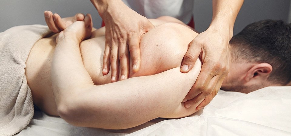
Manual Medicine provides a functional diagnosis of pain and dysfunction in the spine and extremity joints — what we call the axial organ — while manual therapy encompasses hands-on treatment methods, applied for more than a century, for treating these functional disorders, including techniques such as manual therapy, manipulation, mobilization, and post-isometric relaxation.
Manual medicine and manual therapy concern the diagnosis, treatment, and prevention of painful locomotor system diseases.
The methods used in treating pain and dysfunction in the spine and extremity joints are manipulation, mobilization, and post-isometric relaxation techniques.
Unlike the classical approach, these are used alongside existing treatments as hands-on applications to reduce pain and the stimuli causing it.
The results obtained from manual diagnosis, performed using the manual medicine approach, are used to ensure the patient's active participation and, from the perspective of preventive medicine, to prevent disease.
After findings are identified and a diagnosis is determined using the manual medicine approach, patients may be referred to physiotherapists for soft tissue diseases, neuromuscular diseases, and situations requiring mobilization. Until relatively recently, the effect of manual therapy was supported mainly by the practitioner's experience, technique, and the patient's history. However, toward the end of the last century, animal experiment studies were published that described the anatomical and neurophysiological events observed as a result of manual therapy techniques.
Recent publications have observed that manipulation and mobilization applied to the lower back, in suitable patients following examination, produce a significant reduction in pain intensity (Heymann, Fischer, Herget, Nazlikul, Locher, Bronfort, Chou, George, Pickar, Santilli).
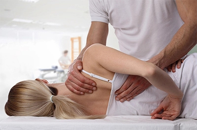 The goal of manual therapy is to increase restricted movement in the joints — referred to as blockage (or dysfunction) — to the highest possible degree, without pain, within postural balance, thereby restoring function and preserving body mechanics.
Safely applying manual therapy requires a detailed anatomical, biomechanical, and neurophysiological examination of the locomotor system.
It should be applied by specially trained specialists who can distinguish indications from contraindications.
Although developments in modern medicine have been dizzying, diseases of the axial organ and spinal system are still far from having a "functional diagnosis."
CT and MRI scans taken for the neck, back, and lower back often fail to explain most patients' clinical complaints.
OUR OBSERVATIONS WITH THE NEURAL THERAPY APPROACH IN MANUAL MEDICINE AND HOLISTIC MEDICINE:
- Pseudoradicular syndrome - 60% (meaning there is no actual nerve root compression)
- Local pain syndrome - 38% (related to local structures such as ligaments, muscles, joints, circulation, etc.)
- Radicular syndrome - 2% (a maximum of about 2% of these problems originate from nerve and nerve root compression)
IT IS WORTH NOTING SOME OF THE STRUCTURES THAT PARTICULARLY CAUSE LOCAL PAIN SYNDROME:
LIGAMENTS
Ligaments are the most important structures contributing to the stability of the axial organ, the spine, and increasing its resistance. Ligaments have inflammatory innervation from the sympathetic nervous system. Contraction or stretching of these ligaments, located in the spine and axial organ regions, causes segmental dysfunction disorders. This both reduces the spine's range of motion and is perceived by the person as pain. These ligament dysfunctions, identified through the manual medicine approach, can be effectively treated by performing stretching exercises with a manual therapy approach while also injecting the affected ligaments with a neural therapy approach at the levels where the dysfunction has been identified.
FACET JOINTS
Facet joints are the joints that guide the movement of a segment and move by gliding. They are covered by synovial tissue and a fibrous capsule. In addition to the ligaments, the facets also make important contributions to the stability of the spine. They also contain receptors that assess the body's position in space (proprioception). They are innervated by the medial branch of the dorsal ramus. Like the ligaments, the facets have a considerable neural innervation and a rich structure in terms of the autonomic nervous system. Facet blockage and degeneration are among the important causes of functional and degenerative restricted movement and pain in the spine.
MUSCLE TISSUE
- The muscle structures located within the local and functional integrity of the vertebrae, the multifidus muscles foremost among them, fundamentally provide the dynamic stability of the axial organ and control of movement. The points where muscles attach to bone are richer in terms of the autonomic nervous system than the muscle itself.
- If the pathological stimuli within a segment cannot be eliminated, the muscles are affected first via the segmental reflex pathway, and increased tone appears.
- A large portion of functional neck, back, and low back pain is muscular in origin, and the most frequently affected muscle is the multifidus. The multifidus is innervated monosegmentally by the dorsal ramus and is responsible for segmental movement. Detecting increased tone in this muscle indicates the presence of segmental dysfunction.
THE FASCIAL CHAIN
- Our body is wrapped in a continuous fascial system extending from head to toe. It is tightly connected to the skin and reaches down to the body's deepest structures. It is connected to all the systems in our body. It has dense nerve receptors, particularly in its superficial layer. It is thought that approximately 2/3 of these nerve fibers are sympathetic fibers. However, because its vascular supply is weak, it cannot repair itself if damaged.
- Thanks to its continuous structure, it functions as a signaling system integrated with the nervous system. Due to its rich innervation, it particularly transmits information from the muscles to distant regions and higher centers. Fascia-related pain is polysegmental. Accordingly, it plays an important role in referred pain.
PAIN AND THE PROTECTIVE MECHANISM
Here, the well-known phenomenon of pain perception and the central nervous system's response to nociceptive stimuli occurs;
At the segmental level, axon collaterals branch off from WDR neurons and reach the alpha and gamma motor neurons to which they belong via interneurons.
As a result, for example, this information reaches the elbow flexor, and the arm moves away from the dangerous area. Simultaneous stopping and position reflexes are stimulated, creating a defense and escape connection. During this process, muscles acting as antagonists are simultaneously inhibited. In other words, to move the arm away from danger, while the biceps muscle contracts, its antagonist, the triceps muscle, is simultaneously inhibited by an inhibitory motor interneuron. These connections form the basis of the protective reflex against danger. Before the brain's conscious protective reaction occurs, the extremities are protected through segmental reflexes.
SEGMENTAL DYSFUNCTION FROM THE MANUAL MEDICINE PERSPECTIVE
If a vertebral joint is overused or misused, nociceptive stimuli related to the state of the extremity travel to the center. Through the protective reflex, spastic protective tension develops in the short autochthonous muscles, the rotators, and the multifidus muscles. When this occurs repeatedly or with high intensity, vertebral dysfunction results from this excessive protective reflex. Blockage, or dysfunction, is, in the manual medicine sense, a form of the protective reflex, and its purpose is to protect the organism.
Such dysfunctions are spatially organized, as in testing joint range of motion, with increased nociceptive input in some directions (the locked direction) and decreased input in others (the free direction). In WDR neurons, it is not only afferents from the relevant joint that converge, but also afferents from various anatomical systems, each originating from a single segment. These are afferents from the skin, muscle, tendon, and internal organs. Nociceptors originating from extravertebral structures — demonstrable through countless clinical examples — cause vertebral dysfunction at the end of the same pathway through activation of the motor system (Heymann 2005, Nazlikul 2010).
Likewise, environmental factors and intrapsychic dispositions also regulate the preparation of motor reactions (the psychomotor pathway). Fear, stress, or a flight response travels through pathways leading to the center and the gamma motor system, priming the muscles and producing increased vertebral joint blockage. What must be done first is to perform a differential diagnosis of the actual problem and resolve it. Generally, the actual problem lies not in the movement system itself, but in nociceptive generators located far from that system. This can sometimes mean that dental issues in the trigeminal region or temporomandibular joint problems are responsible for sacroiliac blockage, or that cervical disc issues are responsible for thoracic blockage (Nazlikul).
In addition, early diagnosis of malignancies should also be considered in the differential diagnosis.
WHAT CAN BE DONE FROM THE PERSPECTIVE OF MANUAL MEDICINE AND MANUAL THERAPY;
- Identifying the problematic segment or segments along the axial organ.
- Using the skin-gliding test to understand the problematic axis and the structures it is, or may be, related to
- Regulating hypo- or hyperlability in the ligaments, which are rich in autonomic nerve supply, particularly sympathetic innervation
- Resolving restricted movement — in other words, blockages — occurring in the facet joint through mobilization and manipulation, and injecting the affected facet, along with neighboring facet joints, with a neural therapy approach
- Manipulation, mobilization, and neural therapy injection of the sacroiliac joint
- Identifying active trigger points, both in the axial organ and in the rest of the body. Injecting these with a neural therapy approach and applying stretching exercises to the relevant muscles
- Evaluating the jaw and the temporomandibular joint (TMJ)
- Assessing and correcting the related organs for walking and posture
Dr. Hüseyin Nazlikul
President of the Manual Medicine Pain Regulation Association (MTAR)
President of the Scientific Neural Therapy Regulation Association
President of IFMANT
Sources Consulted:
• Nazlikul, H: Neural Therapy Textbook
• Nazlikul, H: Neural Therapy — Another Treatment Is Possible
• H. Barop's Neural Therapy Atlas (Translator: H. Nazlikul)
• L. Fischer's Neural Therapy Book (Translators: H. Nazlikul and Y. Tamam)
• James W. McNabb, Joint and Soft Tissue Injections (Translators: H. Nazlikul and Y. Tamam)
• Weinschenk, S: Neuraltherapie
• Fischer, L et al: Lehrbuch Integrative Schmerztherapie